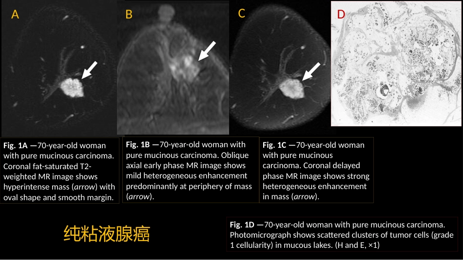
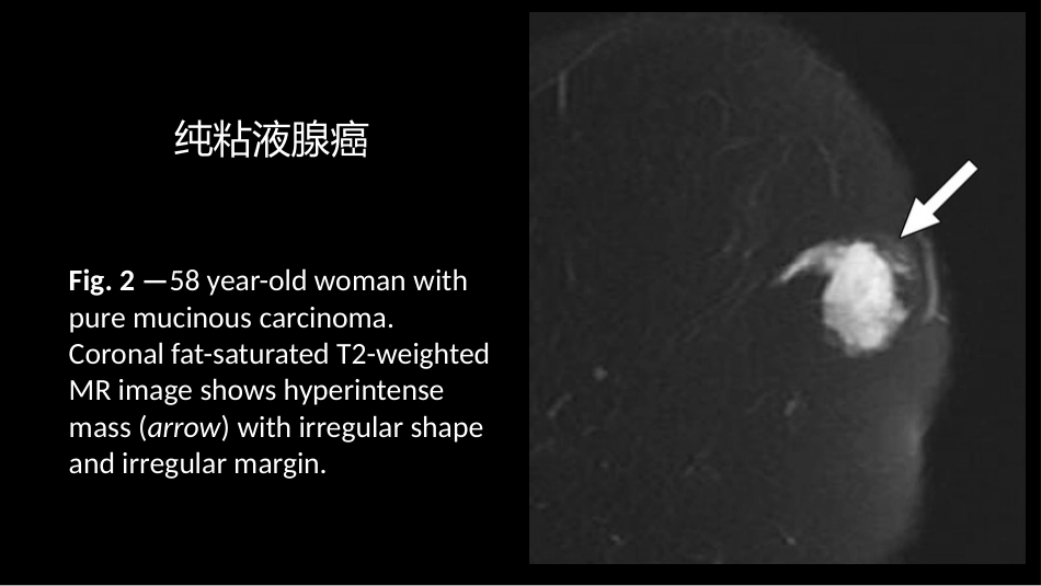
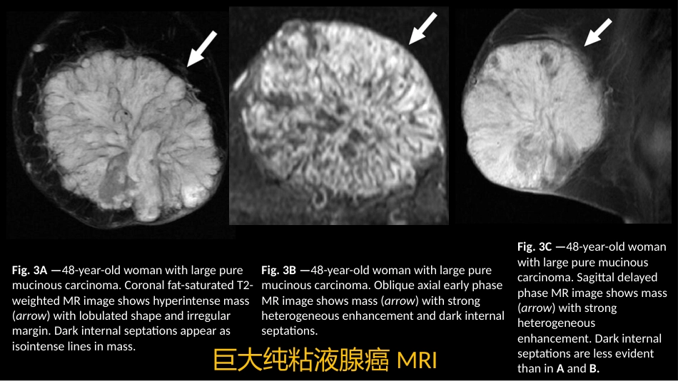
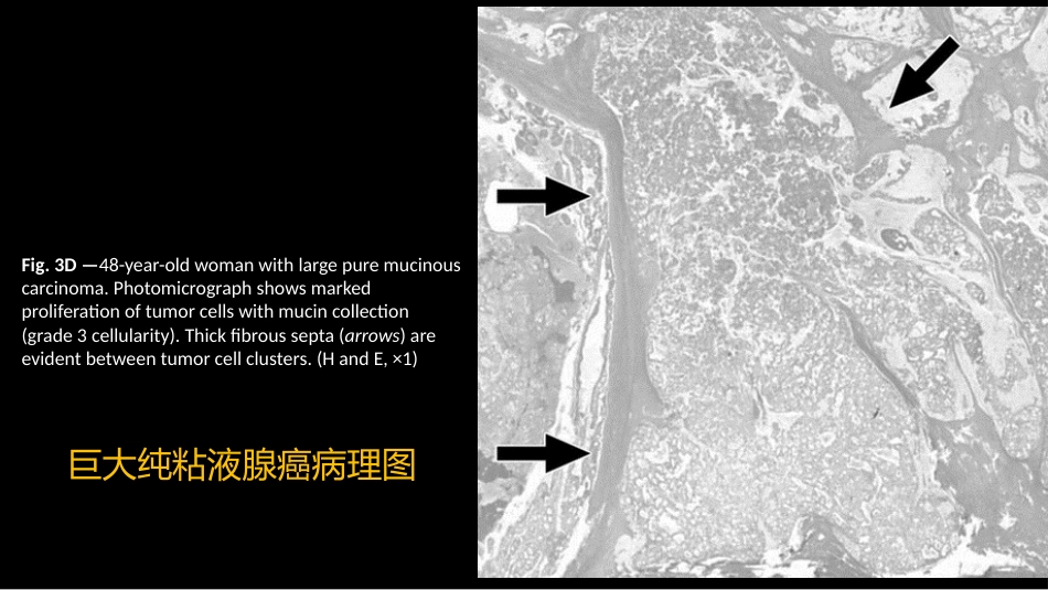
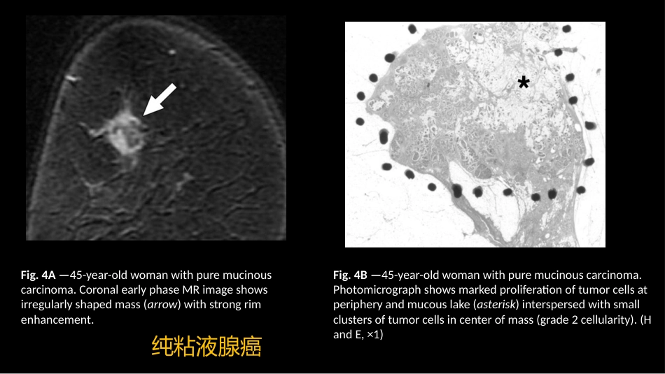
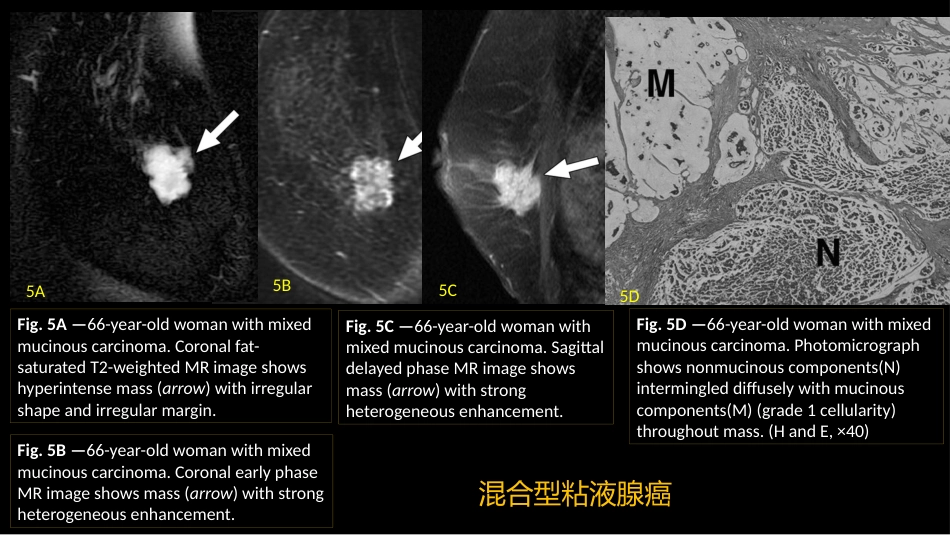
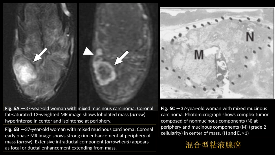
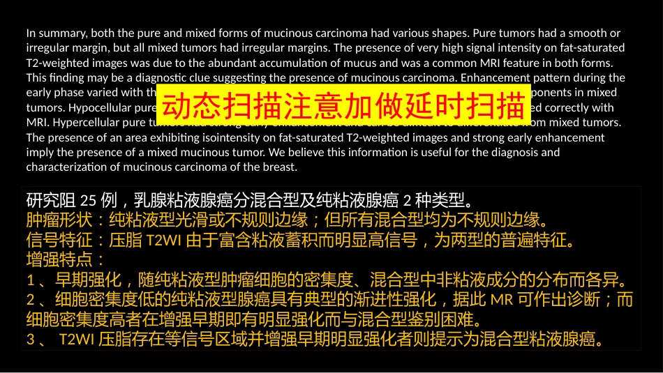
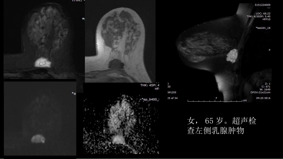
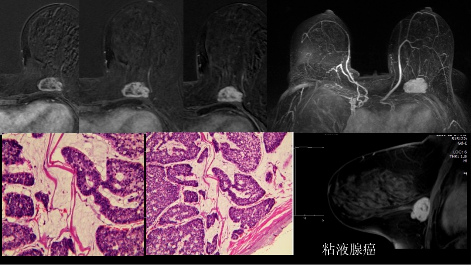
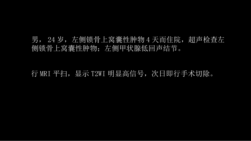
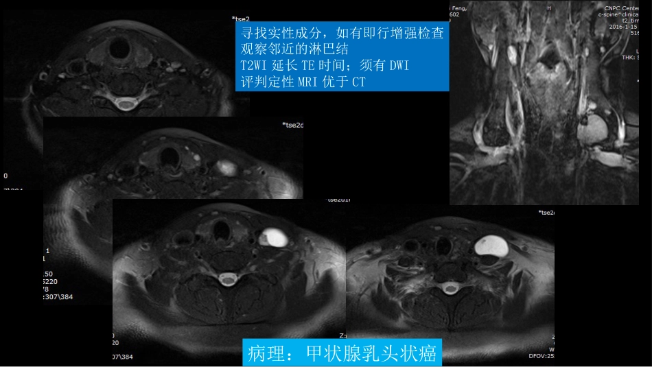
Mucinous Carcinoma of the Breast: MRI Features of Pure and Mixed Forms with Histopathologic CorrelationShuichi Monzawa et al AJR March 2009, VOLUME 192 NUMBER 3乳腺粘液腺癌:纯粘液型、混合型粘液腺癌MRI 特征与病理的关系Fig. 1A —70-year-old woman with pure mucinous carcinoma. Coronal fat-saturated T2-weighted MR image shows hyperintense mass (arrow) with oval shape and smooth margin. Fig. 1B —70-year-old woman with pure mucinous carcinoma. Oblique axial early phase MR image shows mild heterogeneous enhancement predominantly at periphery of mass (arrow). Fig. 1C —70-year-old woman with pure mucinous carcinoma. Coronal delayed phase MR image shows strong heterogeneous enhancement in mass (arrow). Fig. 1D —70-year-old woman with pure mucinous carcinoma. Photomicrograph shows scattered clusters of tumor cells (grade 1 cellularity) in mucous lakes. (H and E, ×1) 纯粘液腺癌ABCDFig. 2 —58 year-old woman with pure mucinous carcinoma. Coronal fat-saturated T2-weighted MR image shows hyperintense mass (arrow) with irregular shape and irregular margin. 纯粘液腺癌Fig. 3A —48-year-old woman with large pure mucinous carcinoma. Coronal fat-saturated T2-weighted MR image shows hyperintense mass (arrow) with lobulated shape and irregular margin. Dark internal septations appear as isointense lines in mass. Fig. 3B —48-year-old woman with large pure mucinous carcinoma. Oblique axial early phase MR image shows mass (arrow) with strong heterogeneous enhancement and dark internal septations. Fig. 3C —48-year-old woman with large pure mucinous carcinoma. Sagittal delayed phase MR image shows mass (arrow) with strong heterogeneous enhancement. Dark internal septations are less evident than in A and B. 巨大纯粘液腺癌MRIFig. 3D —48-year-old woman wi...